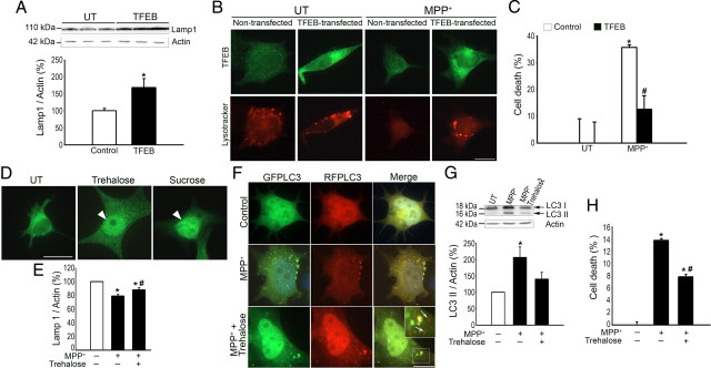
Figure 4.
Enhancement of lysosomal biogenesis by TFEB attenuates MPP+-induced cell death. A, Lamp1 immunoblot levels in control or TFEB-transfected cells. B, LysoTracker fluorescent signal (red) in control or TFEB-transfected cells, UT or intoxicated with MPP+. In transfected cells, TFEB is detected by immunofluorescence (green). C, Cell death in control or TFEB-transfected cells, UT or intoxicated with MPP+, as assessed by MTT assay. D, TFEB immunofluorescence (green) in nontransfected cells, UT or treated with trehalose (1 mm) or sucrose (100 mm) for 24 h. E, Lamp1 immunoblot levels in UT and MPP+-intoxicated nontransfected cells, treated or not with trehalose. F, Fluorescent signal of GFP-LC3 (green) and RFP-LC3 (red) in UT and MPP+-intoxicated tfLC3-transfected cells, treated or not with trehalose (arrows indicate autophagolysosomes, visualized as red-only structures). G, LC3II immunoblot levels in UT and MPP+-intoxicated cells, treated or not with trehalose. H, Cell death in UT and MPP+-intoxicated cells, treated or not with trehalose, as assessed by MTT assay. In all panels, n = 3–5 per experimental group. MPP+, 0.25 mm, unless otherwise indicated. *p < 0.05 compared with UT cells. #p < 0.05 compared with MPP+-treated cells. Scale bar, 10 μm.

