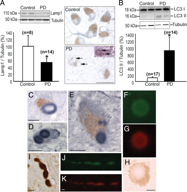
Figure 7.
Lysosomal depletion and AP accumulation in postmortem PD samples. A, Left, Lamp1 immunoblot levels in postmortem substantia nigra protein homogenates from PD patients and age-matched control subjects. A, Right, Lamp1 immunostaining (blue; brown pigment corresponds to neuromelanin) in postmortem substantia nigra sections from PD patients and age-matched control subjects. Arrows indicate Lamp1-negative Lewy bodies, identified by hematoxylin and eosin staining (pink, inset). B, LC3II immunoblot levels in postmortem substantia nigra protein homogenates from PD patients and age-matched control subjects. C–E, LC3-immunoreactive Lewy bodies (blue; brown pigment correspond to neuromelanin) in postmortem substantia nigra sections from PD patients. F–H, Lewy bodies were immunolabeled with both α-synuclein (green, F) and LC3 (red, G). In H, brown pigment corresponds to neuromelanin in transmitted light. I–K, LC3 immunoreactivity was also detected in Lewy neurites in postmortem substantia nigra sections from PD patients (I, LC3 in brown; J, K, LC3 in green, α-synuclein in red). In all panels, *p < 0.05 compared with age-matched control subjects. Scale bars, 10 μm.

