Figure 3.
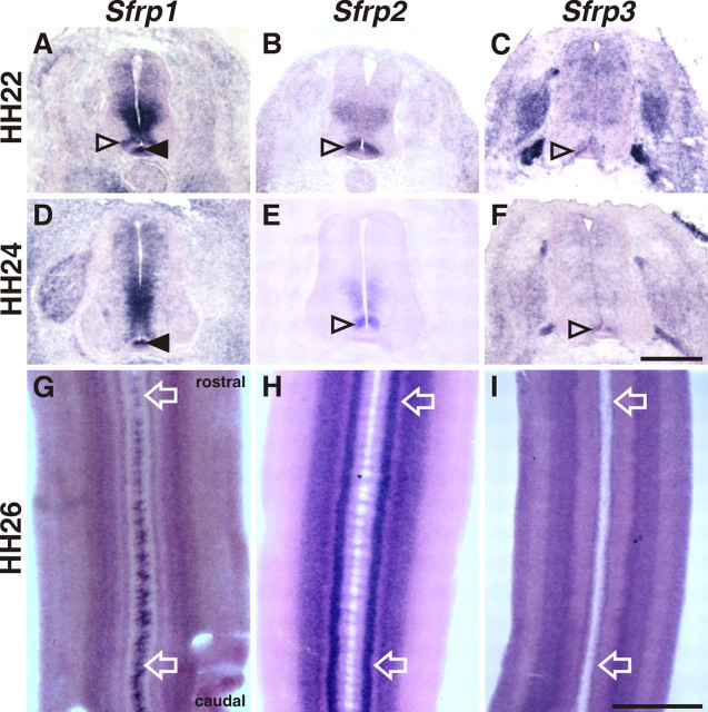
Expression patterns of Sfrps in the embryonic chicken spinal cord. A, D, G, Sfrp1 is expressed in the floor plate (filled arrowhead), in the ventricular zone, and in an area dorsolateral to the floor plate (open arrowhead) at HH22 (A), HH24 (D), and HH26 (G). B, E, H, Sfrp2 expression was found in the ventral ventricular zone with an area of stronger expression dorsal to the floor plate (open arrowhead). C, F, I, Sfrp3 is expressed in an area adjacent to the floor plate (C and F, open arrowheads; I) similar to Wnt7a. In contrast to Wnts, a strong gradient of Sfrp1 was found in the floor plate along the anteroposterior axis with high levels in the caudal floor plate (G) (supplemental Fig. S5, available at www.jneurosci.org as supplemental material). Sfrp2 was expressed in a shallow gradient, in contrast to Sfrp3 that was not expressed in a gradient along the anteroposterior axis. Compare expression indicated by arrows in G–I (supplemental Fig. S5, available at www.jneurosci.org as supplemental material). Rostral is to the top in G–I. Scale bars: (in F) A–F, 200 μm; (in I) G–I, 500 μm.

