Figure 6.
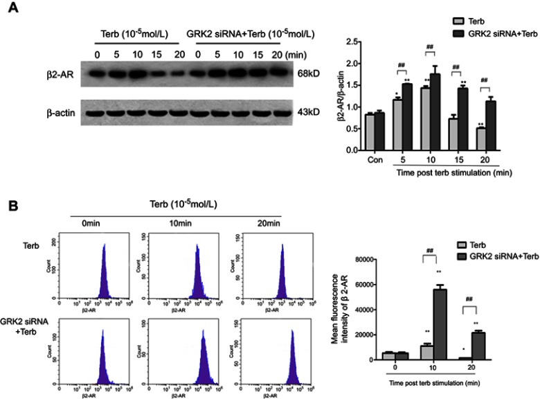
Down-regulation of GRK2 elevated the expression of β2-ARs on the M2-macrophage membrane and delayed the internalization of β2-AR. (A) A Western blot assay was performed to detect the expression of β2-AR on the membrane of M2-polarized macrophages stimulated with Terb for 5, 10, 15 or 20 min following transfection with GRK2 siRNA. The expression of β2-AR on the membrane of M2-polarized macrophages peaked at 10 min in the Terb-alone and GRK2 siRNA+Terb groups. While the expression of β2-AR in the GRK2 siRNA+Terb group was higher compared with that in the Terb-alone group at 10 min. The expression of β2-AR in the Terb-alone group declined rapidly from 10 min to 20 min following Terb stimulation, while in the GRK2 siRNA+Terb group, the expression of β2-AR declined gradually and at 20 min there was still a significant fraction of β2-ARs on the membrane. Histogram represents the relative level of β2-AR in Terb group and GRK2 siRNA+Terb group. (B) Flow cytometry was performed to detect β2-AR expression on the membrane of M2-polarized macrophages stimulated with Terb for 10, or 20 min after transfection with GRK2 siRNA. Though the expression of β2-AR peaked at 10 min in the Terb-alone and GRK2 siRNA+Terb groups, there was a significant elevation in β2-AR formation of the GRK2 siRNA+Terb group compared with Terb-alone group. Furthermore, the decline in β2-AR expression on the cell membrane was slower in the GRK2 siRNA+Terb group compared with Terb-alone group. The histogram represents the relative expression of β2-AR on cell membrane in the Terb-alone and GRK2 siRNA+Terb groups. *P<0.05 and **P<0.01 compared with the 0 min group; ##P<0.01 compared with each other group at the same time point.

