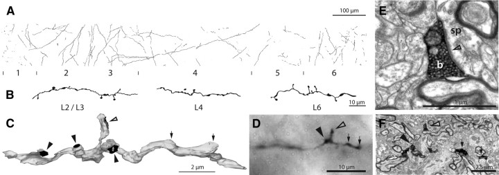
Figure 1.
Innervation of area 17 by claustral axons. A, Light microscopic reconstruction of all the axons innervating a strip of area 17. Laminae and their boundaries are indicated below. B, High-power drawing of three sections of labeled claustral axon taken from different layers. C, Three-dimensional reconstruction from serial ultrathin sections of a labeled claustral axon (gray) in layer 4 showing 3 synapses formed by boutons en passant (solid arrowheads) and 1 synapse from a bouton terminaux (open arrowhead). The two swellings (small arrows) do not contain vesicles and are filled with mitochondria (see also supplemental Fig. 2, available at www.jneurosci.org as supplemental material). D, Light micrograph of corresponding axon (in C) showing the bouton terminaux (open arrowhead), one bouton en passant (solid arrowhead), and the two swellings (small arrows) that could be misleadingly interpreted as synaptic boutons by only considering light micrographs. E, High-power electron micrograph of the vesicle filled bouton terminaux (b) of C and D forming an asymmetric synapse (open arrowhead) with a spine (sp). F, Low-power electron micrograph of identified axon collateral shown in C and D; arrows as in D.

