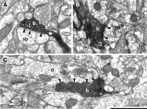
Figure 2.

Electron micrographs of labeled claustral boutons. A, Claustral bouton (b) forming a perforated asymmetric synapse (solid arrowheads) with a spine (sp) in layer 1. B, Claustral bouton (b) forming an asymmetric synapse (solid arrowheads) with a spine (sp) in layer 6. The spine apparatus is clearly visible. C, Claustral bouton forming an asymmetric synapse (solid arrowheads) with a dendrite (d). Scale bar, 1 μm.
