Figure 10.
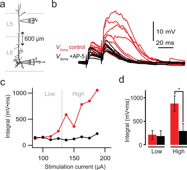
AP-5-sensitive spikes evoked in the distal apical dendrites of L6 pyramidal neurons. a, Experiment setup with a somatic recording electrode (gray) and a distally located extracellular stimulating electrode close (<∼5 μm) to the apical dendrite. Gabazine (0.1 μm) was added to reduce the inhibitory transmission. b, Two extracellularly evoked EPSPs at 50 Hz with progressively increasing stimulus strength up to 200 μA (red traces). AP-5 (50 μm; black traces) blocked the large increase in amplitude and duration of the second EPSP but had only a small effect on the first EPSP. c, Integral of the second EPSP shown in b as a function of stimulus strength. d, Average integral of the second EPSP for five cells. “Low” and “High” refer to response below and above threshold (dashed gray line in c), respectively, for plateau-like responses. Asterisk indicates statistical significance (n = 5; p = 0.013).

