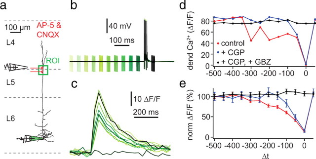
Figure 11.
Activation of distal inhibitory inputs induces a long-lasting blockade of dendritic Ca2+ electrogenesis. a, Experimental arrangement: somatic whole-cell recordings were made with a pipette containing OGB-1 (100 μm; bottom, green) while monitoring ΔF/F at an ROI on the apical dendrite ∼400 μm from soma (green box). An extracellular bipolar electrode was placed in L4 ∼150 μm lateral to the apical dendrite to evoke inhibitory input (represented schematically in red). CNQX (10 μm) and AP-5 (50 μm) were included in the extracellular bathing solution to prevent excitatory synaptic transmission. b, Electrical recordings from the soma (black trace) while evoking a train of three APs at 120 Hz with somatic current injection (2 ms pulses of 1 nA; critical frequency of the cell shown in this example was 84 Hz). The compound IPSP was concurrently evoked by five pulses at 200 Hz. The time of the extracellular stimulation was altered in steps of 50 ms from 500 before to 50 ms after the train of action potentials (green to black traces). c, Gradual blockade of distal Ca2+ transients recorded in the distal ROI. d, Peak amplitudes of ΔF/F as a function of the time interval between the extracellular stimulation and the train of somatic APs (Δt) for the example shown in a–c. Measurements were obtained in control conditions (red) and in the presence of the GABAB antagonist CGP 52432 (1 μm; blue) and after further addition of the GABAA antagonist gabazine (GBZ; 3 μm; black). e, Average inhibition curve for five cells as in d.

