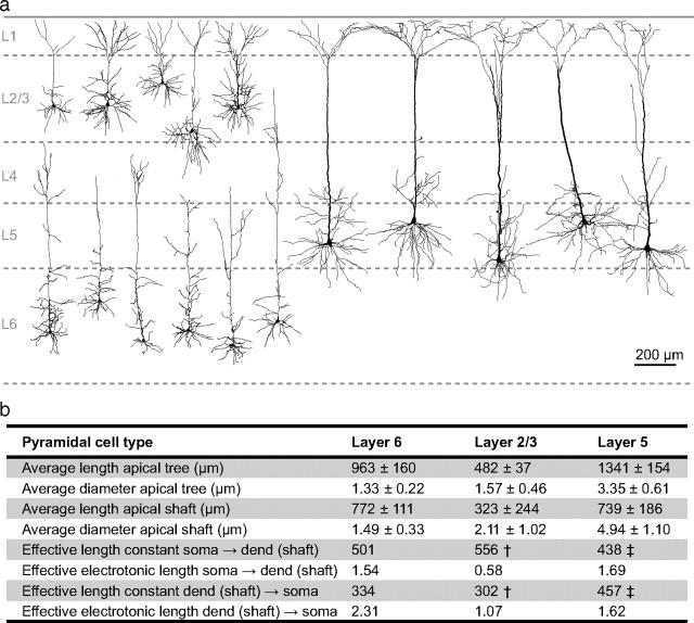
Figure 12.
Anatomical comparison of apical dendrites of different pyramidal neurons in the neocortex. a, Neurolucida reconstructions of pyramidal neurons from layers 2/3, 5, and 6 of the somatosensory cortex in rats. L6 cells were reconstructed from cells used in this study. The L2/3 cells were taken from our unpublished recordings and the L5 cells are those used in the figures of Larkum et al., (2001) for which dendritic and somatic recordings were published (used with permission). b, Table showing the average data for five neurons from L2/3, L5, and L6 neurons from L6 shown in a. Measurements were made for the entire length of the apical tree, i.e., the path from the cell body to the dendritic endpoint furthest from the cell body and also for the apical shaft, i.e., the path from the cell body to the main bifurcation point on the apical dendrite. The terms “effective length constant” and “effective electrotonic length” refer to λeff as defined in the main text and the analogous concept of Leff = l/λeff (where l is the physical length). These are empirically derived distances for the attenuation of steady-state signals traveling along the apical dendrite and should not be confused with the λ and L used for modeling. They are used here for comparison between cell types. †, from Larkum et al. (2007); ‡, from Williams (2004); used with permission.

