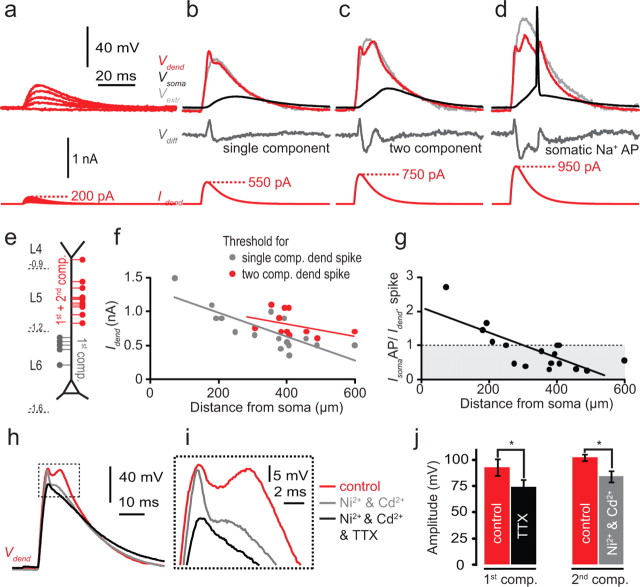
Figure 8.
Dendritic spikes generated by EPSC-like current injection to the dendrite. a–d, Dual recording from the soma and dendrite (at a dendritic distance of 383 μm). Dendritic EPSC waveform current injections (bottom red traces) evoked voltage deflections in the dendrite (top red traces, Vdend) and soma (top black traces, Vsoma). a, Subthreshold EPSC waveform current injection (bottom) evoked similarly shaped voltage deflections in the dendrite (top, red trace). b, Dendritic spike with a fast initial component generated with a peak EPSC amplitude of 550 pA. The extrapolated passive response at suprathreshold EPSC amplitudes is shown in gray in b to d (Vextr). This was used to calculate the active component of the response (Vdiff) by subtracting Vextr from Vdend (middle, dark gray line) c, Dendritic spike with two components generated with increased current injection (750 pA) in the same cell as b. d, Threshold for a somatic AP in the same cell reached with a 950 pA dendritic current injection. e, Diagrammatic representation of the locations of the dendritic current injection that evoked single- and two-component dendritic spikes. The second component could only be generated with current injection more distal than 250 μm from soma, which typically corresponds to the region of the dendrite reaching into L5 and L4. f, Threshold in nA for the single-component (gray circles) and the two-component (red circles) dendritic spike as a function of distance from soma. Linear fits to the data show the progressive decrease in threshold for initiation of single- and two-component dendritic spikes. g, Ratio of the thresholds for the generation of dendritic spikes (Idend) versus somatic APs (IsomaAP) via dendritic current injection. The intersection of the linear fit to the data and the dotted line (IsomaAP/Idend = 1) indicates the approximate position along the apical dendrite where dendritic spikes tend to precede somatic APs or occur in isolation. h, An example of a two-component dendritic spike (red) evoked by EPCS waveform current injection to the dendrite that was reduced to a one-component dendritic spike (gray) by the bath application of Ni2+ (100 μm) and Cd2+ (50 μm). Application of TTX (1 μm; black) blocked the remaining component. i, Enlarged view of the region inside the dashed box in h. j, Average amplitudes of the first and second components before and after application of drugs (n = 3).

