
Figure 7.
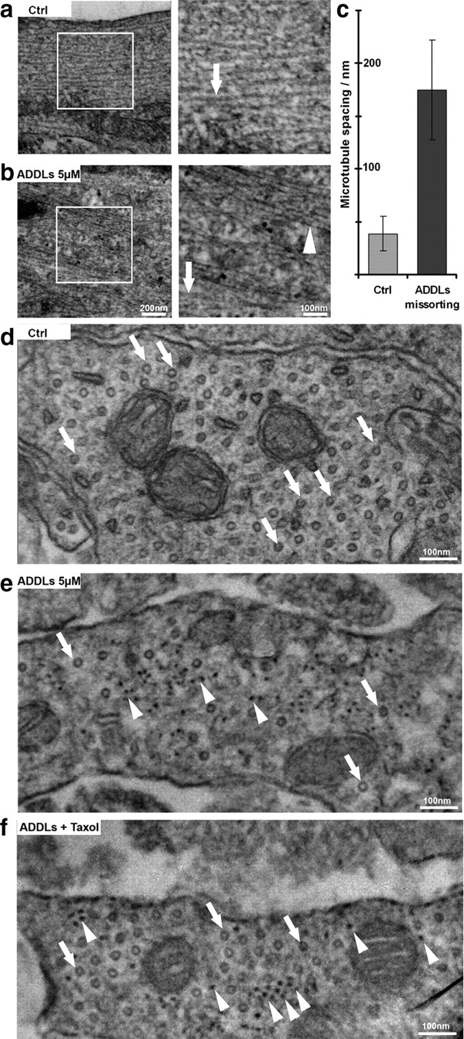
Aβ oligomers induce changes in the cytoskeleton organization. Thin section electron microscopy of dendritic regions of hippocampal neurons (21 DIV) without or with ADDL treatment (5 μm, 3 h). a, Normal cells showing widely spaced microtubules (∼2 diameters apart, arrow) and no neurofilaments. Boxed area on left is shown enlarged on right. b, After ADDL treatment, regions of missorted endogenous Tau (identified by light microscopy after labeling of Tau) contain bundles of neurofilaments (arrowhead) and reduced number of microtubules (arrow). c, Histogram of spacing without or with ADDLs. Error bars: SEM from 4 to 5 cells from 3 independent cultures. d, e, Cross sections of dendrites (d) and dendrites with missorting (e) reveal microtubules as circular shapes (arrows) and dot-like neurofilaments (arrowheads in e). Note the strong decrease in microtubule content in ADDL-induced Tau missorted hippocampal neurons, and the appearance of neurofilaments in e. f, Cross sectioned dendrites after cotreatment with ADDLs and taxol show rescue of microtubules (arrows) and the presence of neurofilaments (arrowheads). Scale bars: a, b left, 200 nm; a, b right; d–f, 100 nm.