Figure 1.

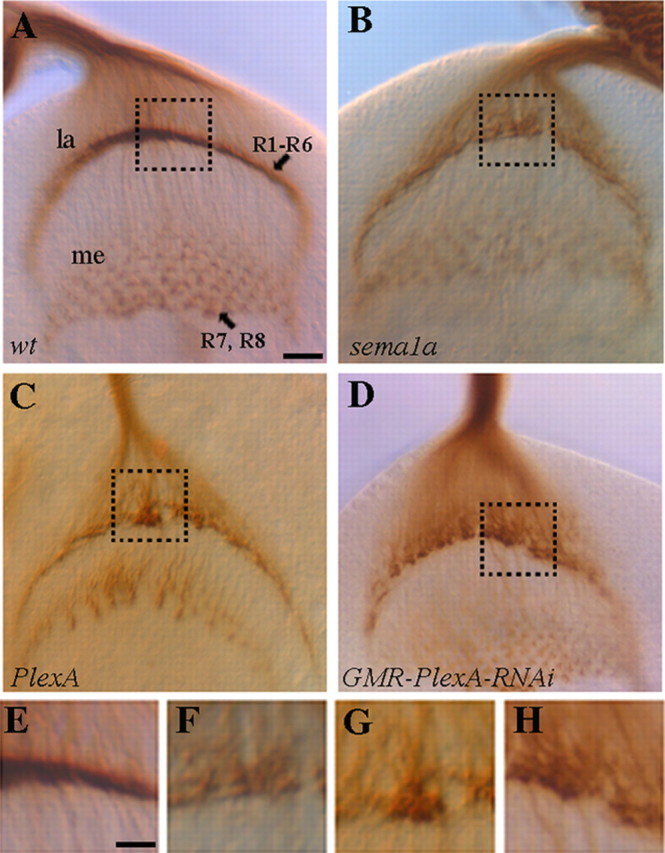
PlexA is required for the proper formation of R1–R6 termination layer in the developing optic lobe. Third-instar larval eye–brain complexes were stained with MAb 24B10 to visualize R-cell axonal projection pattern. A, Wild type. R1–R6 axons stop at the intermediate target region in the lamina (la), where their growth cones expand and associate closely with each other to form a smooth and dense terminal layer. Whereas R7 and R8 axons project through the lamina into the medulla (me). B, sema1aP1 homozygote. R1–R6 terminal layer was disrupted. R1–R6 growth cones associated loosely with neighboring growth cones and failed to pack into a dense layer. C, D, A similar phenotype was observed in PlexA deficiency mutants (C) and eye-specific PlexA knockdown mutants (D). E–H are enlarged views of the boxed regions in A–D, respectively. Scale bar: A–D, 10 μm; E–H, 5 μm.
