
Figure 5.
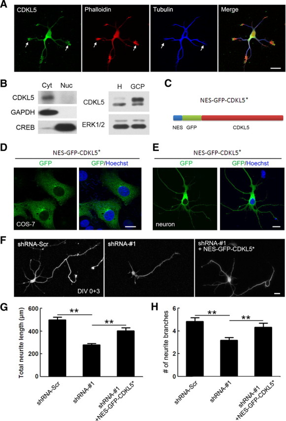
Subcellular localization of CDKL5. A, Representative images of DIV 2 neurons costained with CDKL5 and tubulin antibody. F-actin was visualized by phalloidin staining. Arrows indicate growth cones with abundant CDKL5. Scale bar, 20 μm. B, Left, Western blot showing CDKL5 enrichment in the cytoplasmic fraction, but not the nucleus, of DIV 14 cultured cortical neurons. Cytoplasmic and nuclear lysate were immunoblotted with antibodies against CDKL5, the cytoplasmic factor GAPDH and the nuclear factor CREB. Right, Anti-CDKL5 Western blot analysis of whole-cell homogenate (H) and GCPs isolated from E18 rat cortex. ERK1/2 was used as the loading control. C, Schematic illustration of the protein structure of NES-GFP-CDKL5*. D, E, Representative images showing the localization of NES-GFP-CDKL5* in COS-7 cells (D) and in neurons (E). Scale bar, 10 μm. F, Representative images of neurons transfected with GFP together with the indicated constructs. Scale bar, 20 μm. G, H, Quantitative analysis of total neurite length and total number of dendrite branches in neurons treated as in F. Data represent mean ± SEM; n = 70–80 in each group; **p < 0.001; t test.