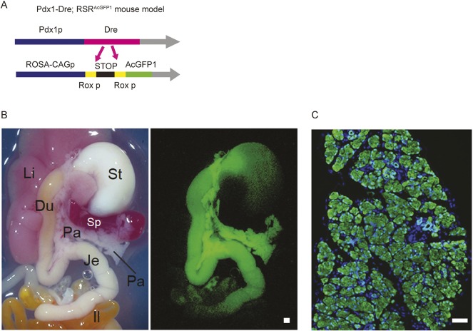
Fig. 2.
Generation of Pdx1-Dre/RSRAcGFP1 mice. (A) Schematic representation of a Dre reporter mouse line (RSRAcGFP1). AcGFP1 is expressed under the control of the CAG promoter after Dre deletes the Rox-flanked stop cassette. (B) Bright-field image (left) and AcGFP1 fluorescent image (right) of Pdx1-Dre/RSRAcGFP1 double-mutant mice at postnatal day 5. AcGFP1 expression is seen in pancreas, antrum of stomach, duodenum and jejunum. Du, duodenum; Il, ileum; Je, jejunum; Li, liver; Pa, pancreas; Sp, spleen; St, stomach. (C) Histological image of Pdx1-Dre/RSRAcGFP1 double mutant mouse pancreas. The section was stained using anti-GFP antibody (green) and a nuclear marker HO (blue). Scale bar: 200 μm.

