Fig. 1.
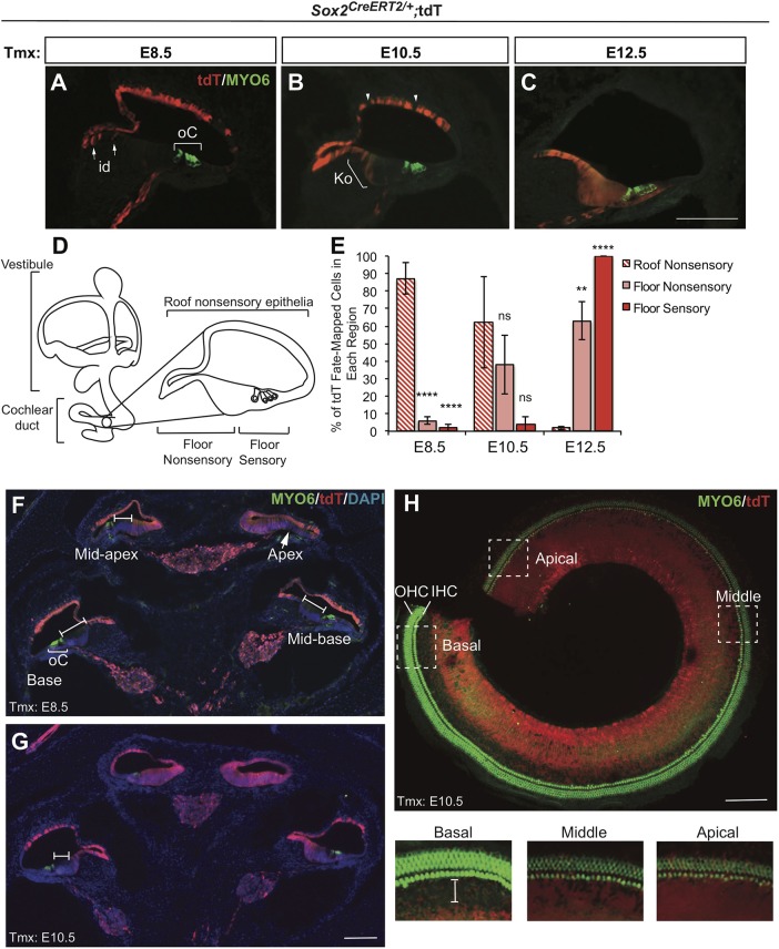
SOX2-expressing cells initially contribute to nonsensory regions in the cochlea but later contribute exclusively to the cochlear floor regions, including organ of Corti. (A-C) Cross-section through the E18.5 cochlea showing tdT/SOX2 expression and hair cell labeling (MYO6). (A) E8.5 tdT/SOX2-expressing cells primarily contributed to the roof of the cochlear duct with the exception of a few interdental (id) cells (arrows). oC, organ of Corti (bracket). (B) E10.5 tdT/SOX2 was downregulated in some cells in the roof of the cochlear duct (arrowheads), and expanded into the floor of the nonsensory cochlea, including the id cell region and some cells in Kölliker's organ (Ko, bracket). (C) By E12.5, expression of tdT/SOX2 was exclusively in the floor of the cochlea, including the oC. (D) Illustration of a cochlea cross-section with respect to the entire inner ear anatomy. (E) The area of nonsensory and sensory regions labeled by tdT/SOX2 was quantified. A trend is apparent, in which SOX2 initially contributes to the nonsensory regions, but switches over time to contribute exclusively to the floor of the cochlea. Comparison of roof nonsensory E8.5 tdT/SOX2 with E8.5 floor nonsensory tdT/SOX2: ****P<0.00001. Comparison of nonsensory E8.5 tdT/SOX2 with E8.5 sensory tdT/SOX2: ****P<0.00001. Comparison of roof nonsensory E12.5 tdT/SOX2 with E12.5 floor nonsensory tdT/SOX2: **P=0.004. Comparison roof nonsensory E12.5 tdT/SOX2 with E12.5 sensory tdT/SOX2: ****P<0.00001. Significance determined using a two-tailed Student's t-test. ns, not significant. Data are mean±s.e.m. (F-G) Midmodiolar regions in Sox2CreERT2/+ E18.5 control cochleae showed that the contribution of tdT/SOX2-expressing cells to the cochlea at E8.5 (F) and E10.5 (G) decreased along the apical–basal axis. At E8.5, tdT/SOX2 was largely excluded from the oC (bracket), with the exception of the apical domain, in which a much smaller negative floor region is observed (arrow) (F). At E10.5, tdT/SOX2 expression expanded along the cochlear floor but remained excluded from the oC in the middle and basal turns (I-bars) (G). (H) Whole-mount cochlea showed E10.5 tdT/SOX2 expression with respect to the sensory region. Boxed areas from different apical and/or basal regions are shown at a higher magnification below. In the apex, tdT/SOX2 was extensively expressed throughout the sensory region, but was gradually excluded more basally (I-bar). IHC, inner hair cell; OHC, outer hair cell. Scale bars: 100 µm.

