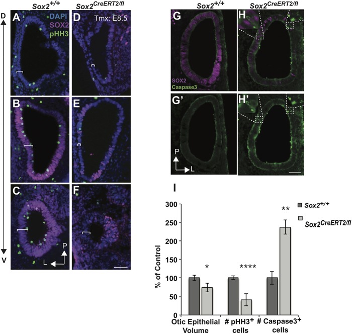
Fig. 10.
SOX2 is required for otic growth and progenitor proliferation and/or viability. (A-F) Sections through E10.5 otocysts of a Sox2+/+ control (A-C) and an E8.5 Sox2CreERT2/fl mutant (D-F) showing a decrease in the proliferation marker pHH3 along the dorsoventral axis (D↔V) in Sox2-deficient mutants. Brackets indicate the thickness of the otic epithelium. (G-H′) The increase in cleaved caspase 3 in the E8.5 Sox2-deleted mutant. Insets show magnification of areas within dashed boxes that display caspase 3-expressing cells in areas where SOX2 was deleted. (I) Quantifications of the reductions in otic epithelium volume (Sox2+/+ n=15; Sox2CreERT2/fl n=12; *P<0.05), pHH3-expressing cells (Sox2+/+ n=9; Sox2CreERT2/fl n=6; ****P<0.0001) and a corresponding increase in caspase 3 (Sox2+/+ n=6; Sox2CreERT2/fl n=6; **P<0.01) in Sox2CreERT2/fl mutants after deletion at E8.5 compared with controls. Significance determined using a two-tailed Student's t-test. Data are mean±s.e.m. L, lateral; P, posterior. Scale bars: 100 µm.

