Fig. 5.
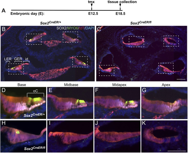
Later deletion of SOX2 severely impairs sensory development in the cochlea but has little impact on cochlear morphology. (A) Time point for deletion (E12.5) and harvest (E18.5). (B) Low power section through the midmodiolar region of a Sox2CreER/+ control at E18.5 shows that E12.5 tdT/SOX2 contributed exclusively to the floor of the cochlear duct in all turns, overlapping with the normal SOX2 protein expression in the supporting cells (white). (C) Section through an E12.5 Sox2-deleted mutant shows relatively normal cochlear morphology but an absence of sensory formation. The missing ganglia in the mutant (tdT labeled in the modiolus in B, marking SOX2-expressing glia at E18.5) probably occurred because of the missing sensory regions, given that it was present earlier at E14.5 (not shown). Magnification of the cochlear turns from the boxed areas in control (D-G) and E12.5-deleted Sox2 mutant (H-K) show the absence of the organ of Corti in all turns, with the exception of an occasional abnormally shaped MYO6-positive cell in the basal turn (H). GER, greater epithelial ridge; id, interdental cell area; LER, lesser epithelial ridge; oC, organ of Corti. Scale bars: 100 µm (in C, representing B,C); 50 µm (K, representing D-K).

