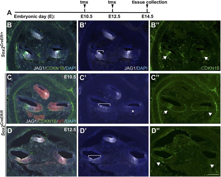
Fig. 6.
Marker analysis in the cochlea indicates that SOX2 is required for both prosensory specification and sensory cell differentiation. (A) Time points for deletion (E10.5 or E12.5) and harvest (E14.5). (B-B″) Control E14.5 midmodiolar region shows typical expression of JAG1 and CDKN1B (marking the future organ of Corti). Unlike JAG1, CDKN1B expression was not always present in control cochlea in the apex. (C-C″) Representative sections of a midmodiolar region of E14.5 cochlea after deletion at E10.5, with indicated prosensory markers and tdT, reflecting SOX2 expression at E10.5. JAG1 expression was mildly expanded in the basal turn of the cochlea (compare brackets in C′ and B′ control) and was significantly downregulated from the middle turn (asterisk) (C′). The shifted JAG1 expression in the apical domain was frequently observed in controls, probably because of a difference in the plane of sectioning rather than a shifted domain. CDKN1B expression was also downregulated in the middle turn (arrowhead) (C″). (D-D″) Representative section of an E14.5 midmodiolar cochlea deleted for SOX2 at E12.5. After deletion of SOX2 at E12.5, JAG1 expression was present in all turns, although, again, mildly expanded in the basal turn of the cochlea (compare bracket in D′ with B′ control). In the absence of SOX2 at E12.5, CDKN1B expression was significantly downregulated in both middle and basal turns (arrowheads) (D″). Arrows and arrowheads indicate either the presence or absence of CDKN1B, respectively. Scale bar: 100 µm.

