Fig. 8.
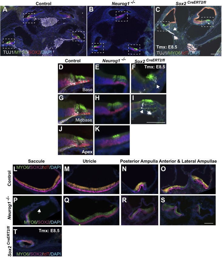
Analysis of NEUROG1-deficient ears indicates that the nonsensory defects are not caused secondarily from reductions in the otic ganglion. (A-C) Low-power midmodiolar sections from control (A), Neurog1−/− (B) and E8.5 Sox2-deleted cochleae (C). The nonsensory phenotype was more severe in the absence of E8.5 SOX2 than of NEUROG1, and some neuronal formation still occurred [arrows mark neuronal expression (TUJ1; white) in C,F,I]. (D-K) Higher magnification views of the boxed regions in A-C highlight that sensory formation occurred fairly normally in the Neurog1-deficient mutant. (L-T) Examples of cross-sections through each vestibular organ in control (L-O), Neurog1−/− mutant (P-S) and E8.5 SOX2-deleted inner ears (T). The vestibule forms relatively normally in Neurog1-deficient mutants, with the exception of a smaller saccule devoid of sensory markers (arrow, P). By contrast, the only vestibular structure seen in E8.5 SOX2-deficient inner ears was an underdeveloped saccule (T). Scale bars: 50 µm.

