
Figure 1.
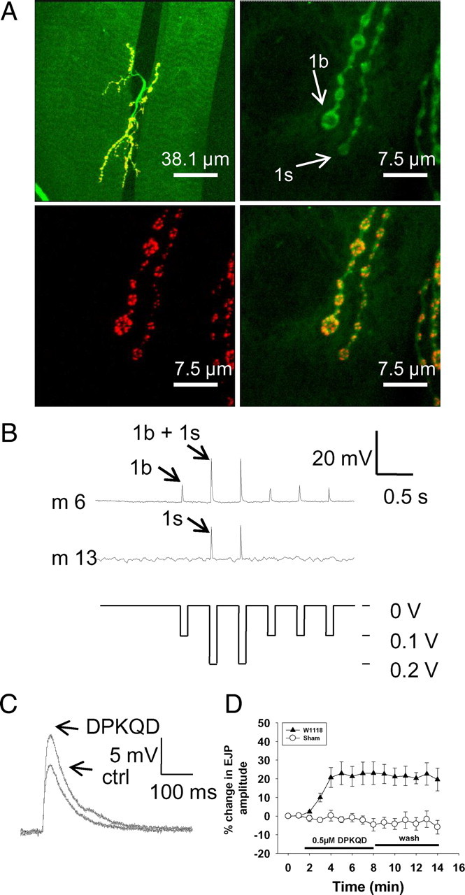
EJP modulation by DPKQDFMRFamide. A, Neuromuscular junctions and synapses of larval Drosophila. At the top left is a low-power view of the neuromuscular junctions on abdominal muscles 6 and 7 formed by two motor axons that supply type 1b and 1s innervation. The boutons are stained with anti-horseradish peroxidase antibody (top right, green) and antibody nc82 against the active zone structural protein Bruchpilot (bottom left, red) to illustrate multiple synapses on each bouton; the merged image is also shown (bottom right). B, Excitatory junctional potentials recorded intracellularly from abdominal muscles 6 and 13. A stimulus intensity of 0.1 V initially recruits the MN6/7–1b (RP3) axon, which innervates only muscle 6 and 7 with tonic-like type 1b nerve terminals. Increase in stimulus intensity to 0.2 V additionally recruits the MNSNb/d-1s axon, which innervates muscles 6, 7, 12, and13 with phasic-like type 1s terminals. Muscle 13 was therefore used as a monitor to detect accidental recruitment of type 1s terminals during the experiment. C, Application of 0.5 μm DPKQDFMRFamide increased the amplitude of EJPs recorded in muscle 6 with stimulation of only type 1b terminals. ctrl, Control. D, Application of 0.5 μm DPKQDFMRFamide induced an increase in amplitude of ∼20% and did not significantly change following washout (wash) of the peptide (n = 6; p = 0.7). “Sham” indicates application of hemolymph-like solution containing no peptide.