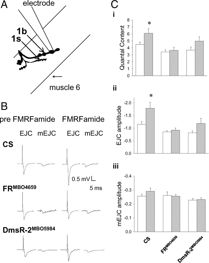
Figure 2.
Modulation of excitatory junctional current recordings from type 1b boutons disrupted in FR but not DmsR-2 receptor mutants. A, Schematic of experimental set-up used to record quantal synaptic currents from type 1b nerve terminals on muscle 6 of the Drosophila abdominal wall. B, Traces of evoked EJCs and mEJCs before and after application of peptide. Ci, Quantal content in CS preparations (n = 6) increased significantly following peptide application from 4.5 ± 0.3 to 6.1 ± 0.6 (two-way ANOVA, p > 0.05), but there was no significant effect of peptide application in either FRMBO4659 (n = 6; p = 0.7) or DmsR MBO5984 (n = 8; p = 0.06) preparations. Cii, The amplitude of EJCs in CS preparations (n = 6) increased significantly from −1.15 mV ± 0.1 mV to −1.8 ± 0.2 mV following peptide application (two-way ANOVA, p > 0.05), but the peptide had no significant effect in FRMBO4659 (n = 6; p = 0.7) or DmsRMBO5984 (n = 8; p = 0.06) preparations. Ciii, The amplitude of spontaneously released mEJCs, measures of postsynaptic sensitivity, did not significantly change following peptide application in CS (n = 6), FRMBO4659 (n = 6), or DmsRMBO5984 (n = 8) preparations (two-way ANOVA, p = 0.4). White bars are before the peptide was added and grey bars are following DPKQDFMRFamide application. Asterisks (*) indicate significant difference.

