
Figure 5.
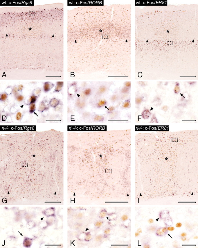
Colocalization of individual laminar markers and c-Fos in wild-type (wt) and reeler (rl−/−) animals. A–L, Stimulated and unstimulated columns in the wild-type cortex (A–F) and reeler cortex (G–L) sectioned in the standard coronal plane, immunostained for c-Fos (brown) and in situ hybridized for different laminar fate markers (purple). The center of the micrographs (A–C) show stimulated barrel-related columns. Activated barrels are marked by stars, and unstimulated barrels are marked by arrowheads at the lamina IV/Va border. Micrographs (G–I) show the corresponding area in reeler with the same labeling conventions. D–F and J–K show higher magnifications of the area indicated by the frames in the micrographs above. Arrows indicate cells that show colocalization of an individual laminar marker and c-Fos. Arrowheads indicate cells singly positive for one of the laminar markers. Scale bars: A–C, G–I, 250 μm; D–F, J–L, 20 μm.