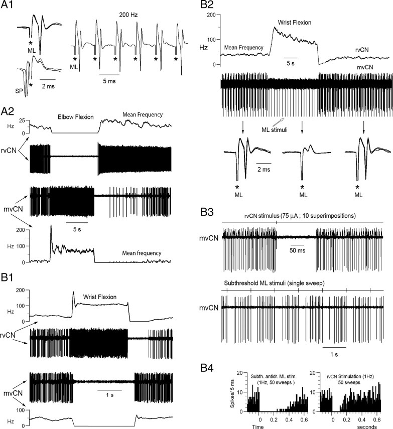
Figure 6.
When simultaneously recorded mvCN and rvCN neurons had receptive fields in distinct articulations, stimulation at the rvCN site inhibited the concurrently recorded mvCN cell. A, B, Two different cells. A, Antidromic identification of a CL neuron (A1) activated by elbow flexion (A2, lower). Elbow flexion inhibited another concurrently recorded rvCN cell (A2, upper). B, Wrist flexion activated an rvCN neuron and inhibited a simultaneously recorded mvCN-CL (B1). The antidromic spikes of the CL cell failed during wrist flexion (B2, superimposed sweeps expanded below), indicating that wrist flexion exerted a true inhibition on the mvCN neuron. Microstimulation at the rvCN recording site transitorily suppressed the activity of the mvCN cell (B3, upper). Subthreshold ML stimulation also transiently silenced the mvCN neuron (B3, lower). The peristimulus histograms in B4 (stimuli at time 0) show that the ML induced inhibition was longer lasting than the rvCN inhibition. Stimulus artifacts are marked with asterisks in A1 and the expanded records in B2; the stimulus artifacts in B3 were recorded on a different channel shown above each neuronal recording.

