
Figure 5.
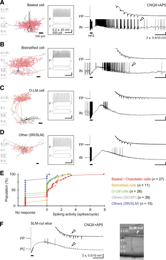
Morphological identification of interneurons showing the prototypic afterdischarge. A, A basket cell showing oscillatory depolarizing responses with robust spiking (arrowhead) during the prototypic afterdischarge in the field potential. Left, Morphology of the recorded and visualized neuron (black, soma and dendrites; red, axons). Middle, Membrane potential responses to depolarizing and hyperpolarizing current injections. Right, Field potential and membrane potential traces during the prototypic afterdischarge. Calibrations are the same in A–D. B, A bistratified cell showing oscillatory depolarizing responses with spiking during the prototypic afterdischarge (arrowhead). These two interneurons were categorized as FS interneurons. C, An O-LM cell showing oscillatory depolarizing responses without spiking during the prototypic afterdischarge. D, Another type of interneuron located in the s. radiatum close to the s. lacunosum moleculare showing no oscillatory responses during the prototypic afterdischarge. E, Cumulative distribution of spiking activity in the prototypic afterdischarge cycles (zero means nonspiking oscillatory responses) in different groups of morphologically visualized interneurons. F, Preservation of the prototypic afterdischarge generation (left, arrowheads) without the s. lacunosum moleculare (right). s.l.m., s. lacunosum moleculare; s.r., s. radiatum; s.p., s. pyramidale; s.o., s. oriens.