
Figure 1.
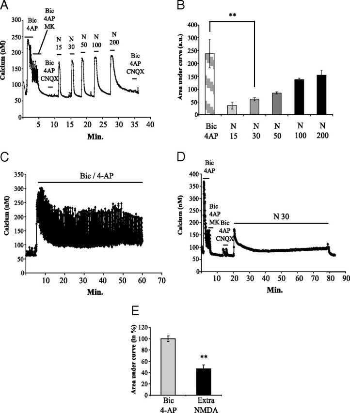
Characterization of prolonged synaptic and extrasynaptic NMDAR activation in cortical neuron cultures at DIV12. Intracellular Ca2+ concentrations were monitored by Fura-2 calcium imaging. A, Cortical neurons were exposed for 2 min to Bic/4-AP and consecutively to Bic/4-AP with MK-801 (10 μm), an open channel blocker, for 3 min to irreversibly block activated synaptic NMDAR. Efficiency of NMDAR blockade was controlled by the absence of Ca2+ response under Bic/4-AP treatment in the presence of CNQX (10 μm), to avoid any AMPA receptor-dependent response. After washing, a dose ranging of NMDA (from 15 to 200 μm for 2 min each) was applied to the culture, and Ca2+ entry in neurons was monitored. Blockade of synaptic NMDAR was finally checked by applying Bic/4-AP + CNQX. B, Calcium responses after Bic/4-AP or increasing doses of NMDA application were quantified by measuring area under curve and represented in histograms (n = 3; n = 137; **p < 0.01). C, Cortical neuron cultures were exposed for 1 h to 50 μm Bic/2.5 mm 4-AP, and cellular Ca2+ entry was monitored using Fura-2. D, After blockade of synaptic response as described in A, cortical neuron cultures were treated for 1 h with 30 μm NMDA to selectively activate extrasynaptic NMDAR. Calcium response was monitored using Fura-2. E, Intracellular calcium concentration was quantified by measuring the area under the curve during the whole treatment duration (1 h for both treatments) (C, D). Statistical analysis was realized by ANOVA followed by Bonferroni–Dunn's test (Bic/4-AP: n = 3; n = 99; 30 μm NMDA after MK801 blockade: n = 3; n = 135; **p < 0.01).