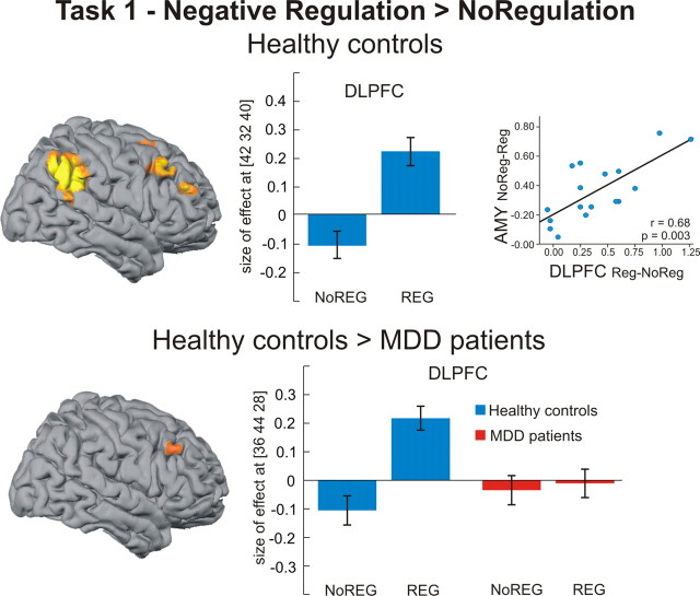
Figure 2.
Regional brain activation during active regulation (task 1). Healthy controls exhibited significant activation of the DLPFC and inferior parietal cortex during active regulation of negative emotions (p < 0.05, family-wise error corrected for whole brain). The activation increase in the right DLPFC was positively correlated with the amount of downregulation in the right amygdala (r = 0.68, p = 0.003, two-tailed). Compared with healthy controls, MDD patients showed reduced DLPFC activation during regulation, as seen in a significant group-by-regulation interaction (p < 0.05, family-wise error corrected for ROI). Bar plots indicate size of effect at the maximum activated voxel in right DLPFC for the contrast negative regulation > no regulation (healthy controls) and the group-by-regulation contrast [healthy controls (regulation > no regulation) > MDD patients (regulation > no regulation)]. AMY, Amygdala; NoREG, no regulation condition; REG, regulation condition.

