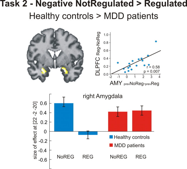
Figure 4.
Regional brain activation during passive viewing (task 2). Healthy controls exhibited a significant sustained regulation effect in the bilateral amygdala. (Note that there was no activation in DLPFC or IPL during task 2 even when the threshold was lowered to an uncorrected p < 0.05). The amount of this sustained regulation effect in the amygdala was positively correlated with DLPFC activation during active regulation in task 1 (r = 0.058, p = 0.007, two-tailed). Compared with healthy controls, MDD patients did not show a sustained regulation effect in the amygdala, as seen in a significant group-by-regulation interaction (p < 0.05 family-wise error corrected for ROI). Bar plots indicate size of the effect at the maximum activated voxel in the right amygdala for the group by regulation contrast [healthy controls (no regulation > regulation) > MDD patients (no regulation > regulation)]. AMY, Amygdala; NoREG, no regulation during task 1; REG, regulation during task 1; prev, previous.

