Figure 1.
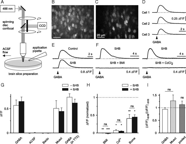
GABA-mediated Ca2+ transients persist in the presence of dl-β-hydroxybutyrate. A, Schematic drawing of the experimental arrangement. B, Raw fluorescence image displaying OGB1-stained cells in the upper cortical plate. C, ΔF image to illustrate GABA-responsive cells (field of view as in B). D, Single-cell somatic intracellular [Ca2+] responses to puff application of GABA (100 μm, 200 ms) (arrowhead). E, βHB (4 mm) did not affect GABA-induced [Ca2+] transients in the presence of TTX. F, GABA-induced [Ca2+] transients were sensitive to antagonists of GABAA receptors (BMI, 100 μm) and voltage-gated Ca2+ channels (CdCl2, 100 μm). Examples in E and F represent averages of eight to ten cells. G–I, Quantification of results. G, Pooled data from unpaired and paired experiments. H, Significance levels for each group were calculated using one-sided paired t tests (BMI/−βHB, n = 5; BMI/+βHB, n = 5; Cd2+/−βHB, n = 4; Cd2+/+βHB, n = 5; bumetanide/−βHB, n = 7; bumetanide/+βHB, n = 5). Baclo, Baclofen; Musci, muscimol; Bume, bumetanide. ns, Not significant; result of a two-way ANOVA in H and result of a paired t test in I. *p < 0.05, **p < 0.01, ***p < 0.001.

