
Figure 3.
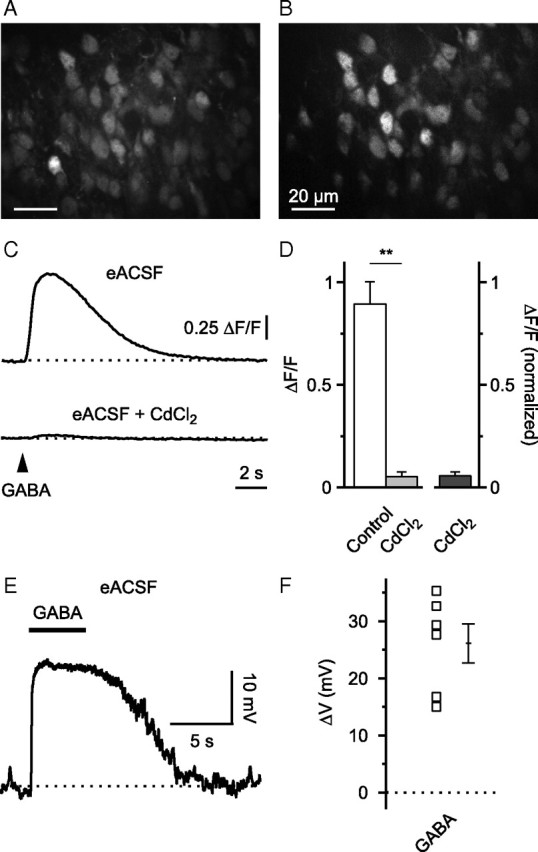
GABA-mediated somatic Ca2+ transients and depolarization persist in energy-substrate enriched ACSF. A, Raw fluorescence image displaying OGB1-stained cells in the upper cortical plate. B, ΔF image to illustrate GABA-responsive cells. C, In eACSF, GABA-mediated somatic [Ca2+] transients were sensitive to CdCl2 (100 μm). Sample traces are averages of five cells. D, Quantification of results. E, Cell-attached current-clamp recording. Puff application of GABA (100 μm) induced membrane depolarization (action potentials blocked by TTX). F, Quantification of results. **p < 0.01.