
Figure 4.
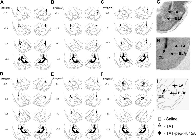
Cannula placements. A, B, Cannula tip placements from rats injected with saline, TAT peptide or TAT-pep-R845A peptide before training tested for LTM (A) or STM (B). C–E, Cannula tip placements from rats injected with TAT peptide or TAT-pep-R845A peptide immediately after training (C), before LTM test (D) or 24 h after training tested 48 h later (E). F, Cannula tip placements from rats injected with TAT peptide, TAT-pep-R845A peptide or saline into CE before training and tested for LTM. G, H, Photomicrograph of injecting cannula trace LA (G) or CE (H). I, Schematic drawing of amygdala nuclei.