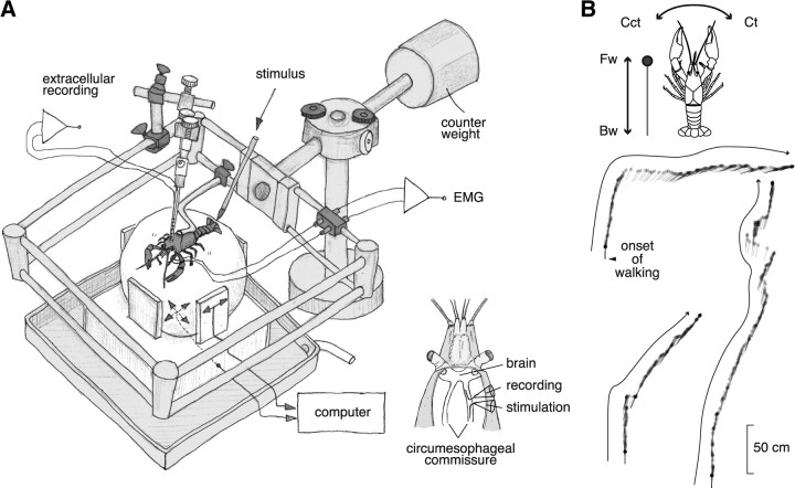
Figure 1.
A spherical treadmill system used for extracellular recording from the brain of crayfish during walking and its quantification. A, An overview of the system. Two optical mice were placed on the basal and side planes. Those mice translated the three rotation vectors of the sphere movement caused by the spontaneous or mechanical stimulus-evoked walking of crayfish. The animal could stand up and down freely on the sphere together with the electrode and its holder whose weight was canceled out by the counter weight. A tactile stimulus was given to the telson manually with a soft brush. For defining the behavioral onset time, we made electromyographic recordings from the mero-carpopodite flexor muscle in the second walking leg on either side (Chikamoto et al., 2008). The inset shows a schematic dorsal view of the recording and stimulation with suction electrodes from the cut ends of small bundles isolated from the circumesophageal commissure. B, Behavioral data of the animal during the recording session. The animal showed forward walking three times. The filled circle indicates the animal head, and the straight line indicates the body axis orientation. The trajectory consists of schematic representation of the animal body sampled every one-sixth s. Fw, Forward walking; Bw, backward walking; Ct, clockwise turning; Cct, counterclockwise turning.

