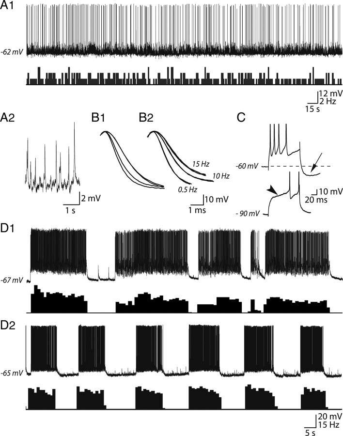
Figure 2.
Basal electrical properties of VP neurons in organotypic slice cultures. A1, The top trace shows an intracellular recording from a VP neuron in a 10-week-old culture from a 9-d-old rat. The neuron displayed a slow irregular activity varying from 0.2 to 3 Hz as indicated by the sequential distribution histogram of AP discharge (bottom trace). In addition to APs, a sustained synaptic activity was clearly visible (A2). B, Example showing the progressive duration increase (broadening) of APs during a burst (B1). The broadening was also frequency dependent as illustrated in B2 in which APs were triggered by positive current pulses (+50 pA, 50 ms; in the presence of CNQX) at increasing frequencies (0.5, 10, and 15 Hz). C, Injection of a depolarizing current pulse (+70 pA, 100 ms) (data not shown) applied at resting potential (−60 mV) triggered a burst of APs. Note the presence of an afterhyperpolarizing potential (AHP) after the train of APs (arrow). When the cell was hyperpolarized to −90 mV, a notch (arrowhead) delayed the occurrence of APs. D, Two examples of spontaneous phasic activity characterized by successive bursts of APs and silent periods (D1, D2, top traces). The bottom traces are the corresponding sequential distribution histogram of APs, typical of phasic activity. Note the synaptic activity visible between bursts in D2.

