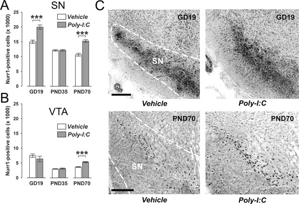
Figure 2.
Age-dependent alterations in mesencephalic expression of the orphan nuclear transcription factor Nurr1 following prenatal immune challenge. Pregnant mice were exposed to the viral mimic Poly-I:C or vehicle treatment, and the effects on Nurr1 protein expression in midbrain structures were investigated in the resulting offspring at the fetal (GD19), peripubertal (PND35), and adult (PND70) stages of development using stereological estimations of Nurr1-positive cells on serial coronal brain sections. A, Maternal Poly-I:C treatment significantly increased the number of Nurr1-positive cells in the fetal SN relative to maternal vehicle treatment. At the peripubertal stage of development, prenatally immune challenged and control offspring did not differ in the number of Nurr1-positive neurons in the SN region. However, a significant increase in the number of SN Nurr1-positive cells was noticeable in adult offspring born to Poly-I:C-challenged mothers in comparison with age-matched control offspring. ***p < 0.001, based on Fisher's LSD post hoc group comparisons of GD19 or PND70 specimen following the presence of a significant two-way interaction in the initial 2 × 3 (prenatal treatment × age) ANOVA (F(2,63) = 17.38, p < 0.001). B, Prenatal Poly-I:C exposure led to a significant increase in Nurr1-positive cells in the VTA specifically in adult (but not peripubertal or fetal) offspring compared with adult offspring of control mothers. ***p < 0.001, based on Fisher's LSD post hoc group comparisons of PND70 specimen following the presence of a significant two-way interaction in the initial 2 × 3 (prenatal treatment × age) ANOVA (F(2,63) = 4.06, p < 0.05). C, Representative images of coronal brain sections of fetal (GD19) and adult (PND70) offspring derived from vehicle- or Poly-I:C-treated mothers stained for Nurr1 protein by immunohistochemistry. The images show Nurr1 cell stainings in the fetal SN and VTA, and in the adult SN. For all developmental stages, Nurr1-positive cells in the midbrain were clearly identifiable by the appearance of darkly stained cell bodies. Scale bars, 250 μm. All values in A and B are means ± SEM. The numbers of offspring included in the analyses were N(GD19-vehicle) = 12, N(GD19-Poly-I:C) = 11, N(PND35-vehicle) = 12, N(PND35-Poly-I:C) = 11, N(PND70-vehicle) = 11, N(PND70-Poly-I:C) = 12.

