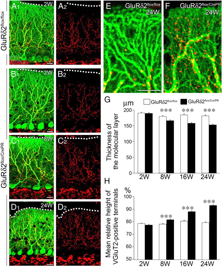
Figure 1.

Progressive atrophy of the distal dendrites and molecular layer of PC with reciprocal expansion of CF territory after induction of GluRδ2 ablation. A–F, Double immunofluorescence for calbindin (green) and vesicular glutamate transporter VGluT2 (red) in GluRδ2flox/flox at 2 weeks after RU-486 administration (A) and GluRδ2flox/CrePR mice at 2 weeks (B), 8 weeks (C), and 24 weeks (D) after RU-486 administration. A2–D2 are separated images of A1–D1, respectively. Note atrophied distal dendrites of PCs and aberrant distal extension of CF innervation in GluRδ2flox/CrePR mice at 24 weeks after RU-486 administration (F), compared with normal territorized innervation in GluRδ2flox/flox mice (E). G, Changes in the thickness of the molecular layer after RU-486 administration. The mean thickness (in micrometers) is 191.0 ± 2.0 (n = 31 sites), 180.1 ± 2.6 (n = 43), 184.7 ± 3.6 (n = 21), and 181.9 ± 2.7 (n = 27) in GluRδ2flox/flox mice, and 189.9 ± 2.3 (n = 47), 166.6 ± 2.3 (n = 49), 158.5 ± 2.2 (n = 44), 153.4 ± 1.8 (n = 31) in GluRδ2flox/CrePR mice at 2, 8, 16, and 24 weeks after RU-486 administration, respectively (mean ± SEM; p = 0.9146 at 2 weeks; p < 0.00001 at 8, 16, and 24 weeks, U test). H, Changes in the vertical height to the tips of VGluT2-positive CF terminals relative to the thickness of the molecular layer. Scores (in percentage) are 78.7 ± 0.4 (n = 49 sites), 78.2 ± 0.4 (n = 49), 80.6 ± 0.6 (n = 29), and 79.4 ± 0.5 (n = 27) in GluRδ2flox/flox mice, and 77.5 ± 0.5 (n = 43), 81.7 ± 0.4 (n = 52), 88.1 ± 0.5 (n = 44), and 93.1 ± 0.3 (n = 31) in GluRδ2flox/CrePR mice at 2, 8, 16, and 24 weeks after RU-486 administration, respectively (mean ± SEM; p = 0.0979 at 2 weeks; p < 0.00001 at 8, 16, and 24 weeks, U test). ***p < 0.001. Scale bars: A–E, 20 μm.
