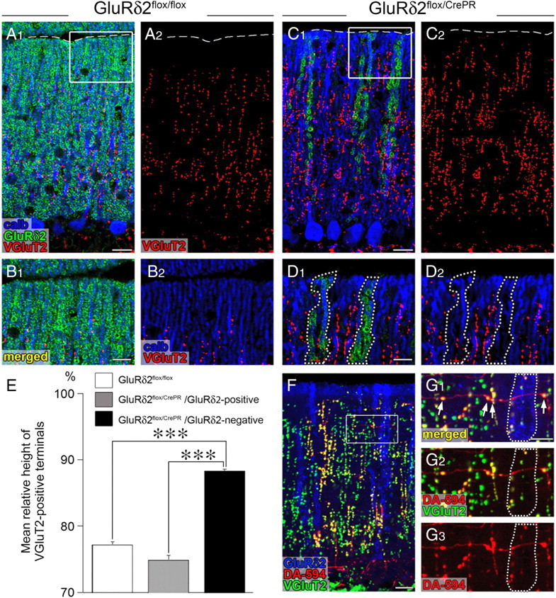
Figure 2.

Aberrant extension and innervation of CFs along and against GluRδ2-negative PC dendrites. All data were obtained using horizontal cerebellar sections of in GluRδ2flox/flox (A, B) and GluRδ2flox/CrePR (C, D, F, G) mice at 12 weeks after RU-486 administration. The boxed regions in A, C, and F are enlarged in B, D, and G, respectively. A–D, Triple immunofluorescence for calbindin (blue), GluRδ2 (green), and VGluT2 (red). Note that the reach of VGluT2-positive CF terminals selectively extends along GluRδ2-lacking dendrites, compared with that along GluRδ2-positive dendrites (encircled by dotted lines in D). The broken lines in A and C indicate the pial surface. E, A histogram showing the mean height to the tips of VGluT2-positive CF terminals relative to the height of the molecular layer. White bar, GluRδ2flox/flox mice; gray bar, GluRδ2flox/CrePR mice (along GluRδ2-positive dendrites); black bar, GluRδ2flox/CrePR mice (along GluRδ2-negative dendrites). Error bars indicate SEM. F, G, Triple fluorescent labeling for GluRδ2 (blue), anterograde tracer DA-594 (red), and VGluT2 (green). Note that VGluT2-positive terminals are preferentially differentiated where transverse CF branches cross with GluRδ2-negative dendrites, whereas such terminal differentiation is rarely found around GluRδ2-positive dendrites (encircled by dotted lines in G). ***p < 0.001. Scale bars: A1, C1, F, 20 μm; B1, D1, G1, 10 μm.
