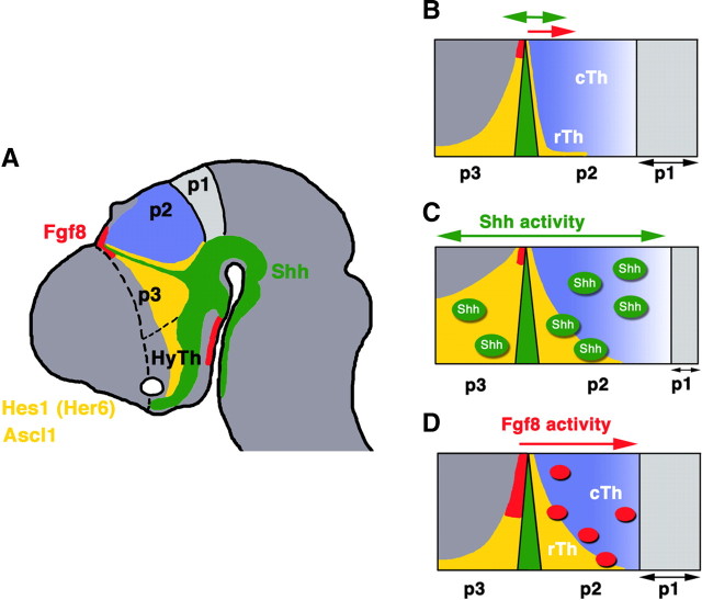
Figure 1.
A, Sagittal schematic of mouse embryonic brain at E10.5. The diencephalic neuraxis is divided in p1, p2, p3, and hypothalamus (posterior to anterior). Presumptive borders of the telencephalon and diencephalon, p3, and hypothalamus are shown by dashed lines. Shh expression domains in basal diencephalon and MDO/Zli are indicated in green, Fgf8-expressing region in dorsal diencephalon and hypothalamus are indicated in red, and Hes1 (Her6) and Ascl1 (acc1) are indicated in yellow. B, Schema of detailed subdivision in the developing thalamus. Expression of Shh in MDO (green) divides p2 and p3, and expression of Fgf8 in dorsal diencephalon is detectable only in p3 (red). The p2 region can be further subdivided in two subregions: rostral thalamus [rTh (or the Rim)]; and caudal thalamus (cTh). Shh signaling is detectable both anterior and posterior to the MDO, whereas Fgf signaling is detectable only posterior to this region. C, Overexpression of Shh expands the Hes1 and Ascl1 in both p2 and p3. D, Overexpression of Fgf8 expands the Hes1 and Ascl1 only in p2.

