
Figure 5.
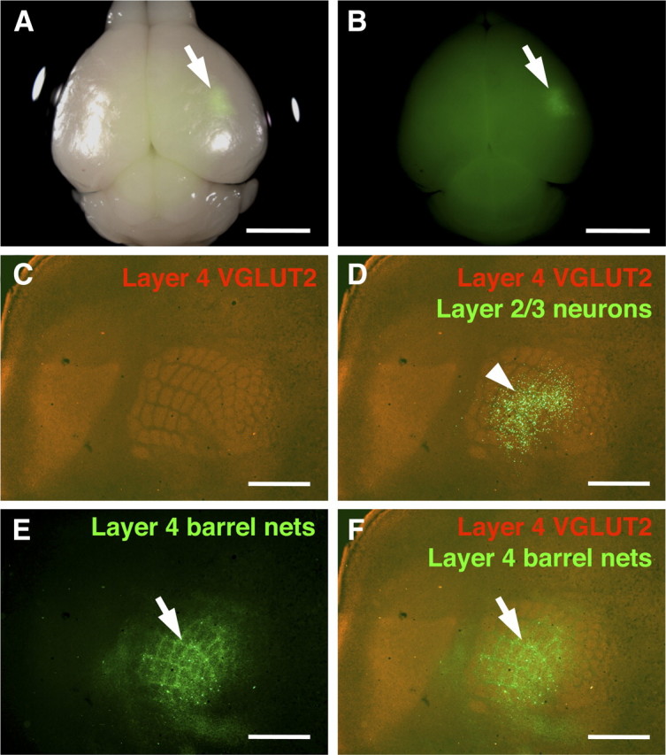
Selective expression of GFP within the barrel cortex using focal in utero electroporation. In utero electroporation was performed at E15.5 with small electrodes of 1 mm diameter, and the brain was dissected at P15. A, A macroscopic image of the brain. A bright-field image and a GFP fluorescence image are merged. GFP signal (arrow) was observed in a small region. Scale bar, 3 mm. B, A single-channel image of GFP fluorescence from A is shown. Arrow indicates a GFP-positive area. Scale bar, 3 mm. C–F, The cortical hemisphere of the electroporated side was flattened, and tangential sections of 50 μm thickness were made. The section containing layer 4 was stained with anti-VGLUT2 antibody. VGLUT2-positive TCAs (C), GFP-positive axons (E), and a merged image (F) are shown. In D, the single-channel image of VGLUT2 staining was also overlaid with a GFP fluorescence image from another section, which contained GFP-positive layer 2/3 neurons, using blood vessels as landmarks. Note that barrel nets are clearly visible (arrows) even when the area containing GFP-positive cells is restricted to the barrel field (arrowhead). Scale bars, 1 mm.