
Figure 7.
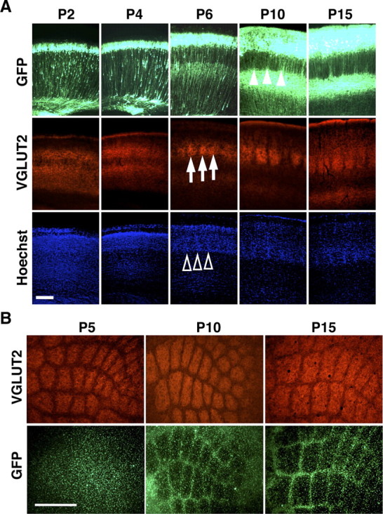
The formation of barrel nets during development. A, Coronal sections of the barrel cortex were prepared at the indicated ages and stained with anti-VGLUT2 antibody and Hoechst 33342. Note that whisker-related patterns of TCAs (arrows) and cytoarchitectonic barrels (open arrowheads) are visible at P6, whereas GFP-positive barrel nets become detectable at P10 (closed arrowheads). Scale bar, 200 μm. B, Tangential sections prepared at indicated ages were stained with anti-VGLUT2 antibody. Note that whisker-related patterns of TCAs are present at P5, while GFP-positive barrel nets are visible only at P10 and P15. Scale bar, 500 μm.