
Figure 2.
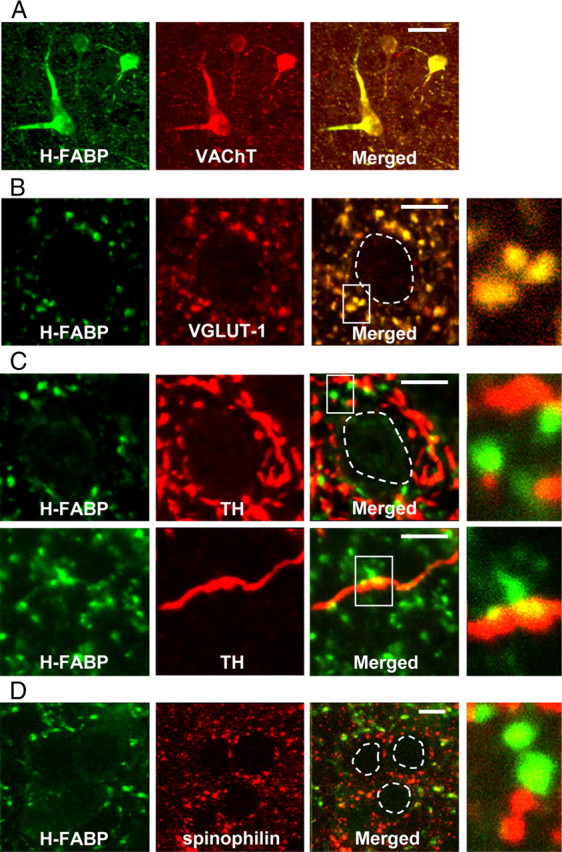
H-FABP localization in the dorsal striatum. Confocal images showing colocalization of H-FABP (green) and markers of three classical neurotransmitters (acetylcholine, glutamate, and dopamine) or spinophilin (red) in the dorsal striatum. A, Immunoreactivities of H-FABP and VAChT almost completely merge. B, Most H-FABP-containing boutons show VGLUT1 immunoreactivity. C, D, Most H-FABP-positive structures do not show immunoreactivity for either TH or spinophilin. At right in B–D are high-magnification images. Areas circled with dashed lines are cell soma. Scale bars: A, 30 μm; B–D, 10 μm.