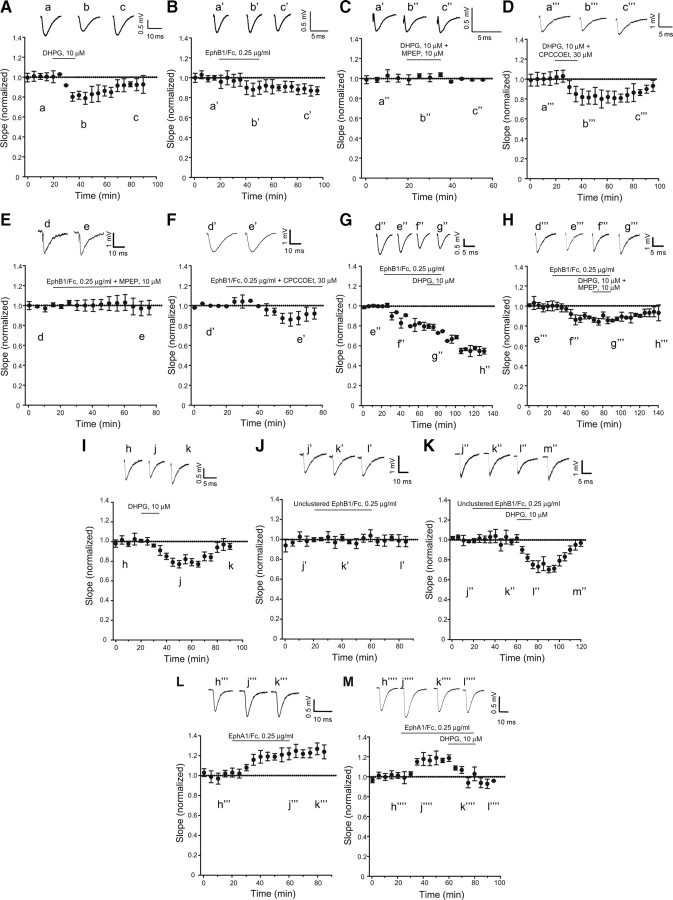
Figure 2.
Same as in Figure 1, but in hippocampal slices from rats at postnatal days 7–9. In A and D (n = 6 and 4, respectively), values corresponding to the peak of synaptic depression (40–60 min) were significantly different from baseline values (p < 0.05); in B (n = 5), values following the termination of Eph/Fc exposure were significantly different from baseline values (p < 0.05); in F (n = 5), values from 50 to 60 min were significantly different from baseline values (p < 0.05); in C and E (n = 4 and 5, respectively), none of the values with DHPG + MPEP or clustered EphB1/Fc + MPEP differed from baseline values; in G (n = 5), we found the same synergism between EphB1/Fc and DHPG observed in adult hippocampal slices (see Fig. 1G). All values recorded at the end of DHPG exposure were significantly different from values recorded during exposure to EphB1/Fc alone (p < 0.05). Again, the synergism between EphB1/Fc and DHPG was abolished when DHPG was applied in the presence of MPEP (n = 5). Pool data from a different set of experiments with hippocampal slices from rats at postnatal days 7–9 are shown in I–M. In I (DHPG; n = 5) and K (unclustered EphB1/Fc + DHPG; n = 5), values corresponding to the peak of synaptic depression (from 40 to 80 min in I and from 79 to 110 min in K) were significantly different from baseline values (p < 0.05); the lack of effect of unclustered EphB1/Fc on synaptic transmission is shown in J (n = 6); in L (n = 6), clustered EphA1/Fc is shown to induce a long-lasting increase in synaptic transmission. Values at times >40 min are significantly different from baseline values; in M (n = 5), DHPG is still able to reduce synaptic transmission in the presence of clustered EphA1/Fc. Representative traces illustrating fEPSP are shown in all figures. Traces are averages of four consecutive responses at the indicated time points.

