Figure 8.
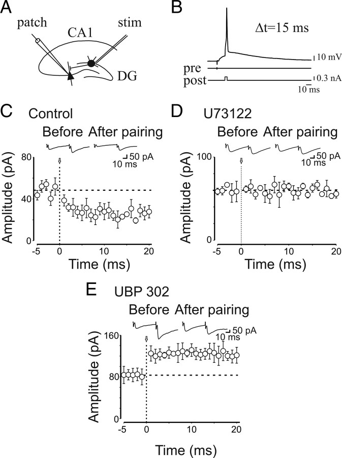
At immature MF–CA3 synapses, presynaptic kainate receptors control the direction of STDP. A, Schematic representation of the experimental design. B, The stimulation of granule cells in the dentate gyrus (pre) preceded the postsynaptic spike (post) by 15 ms (Δt). C, Summary plot of the mean peak amplitude of GPSCs recorded in the presence of GYKI 52466 before and after pairing (arrow at time 0; n = 11). The dashed line represents the mean amplitude of GPSCs before pairing. The insets represent averaged GPSCs obtained from a single neuron before and after pairing. Note that pairing induced synaptic depression. D, Summary plot of MF-GPSCs amplitude obtained in the presence U73122 versus time (n = 7). Note that blocking PLC with U73122 failed to produce any effect on GPSCs amplitude. E, As in C, but in the presence of UBP 302 (n = 9). In this case, pairing induced synaptic potentiation. Error bars indicate SEM.

