
Figure 5.
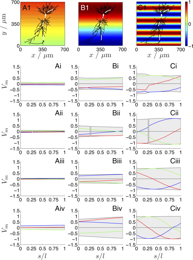
The effect of a spatially harmonic one-dimensional electric field (along the somatodendritic axis) on the Vm of the reconstructed CA1 neuron (top row; white lines indicate the same four sections as in Fig. 4A) for three spatial frequencies: λs = 6.25 mm (fs = 0.16 mm−1; A), 1 mm (1 mm−1; B), and 0.2 mm (5 mm−1; C). The color map illustrates the (spatially) one-dimensional extracellular Ve oscillation along the somatodendritic axis for a phase ϕy. The distance along each section is shown as dimensionless arc length s/l. i is the basal; ii, the soma and the proximal apical; iii, the medial apical; and iv, the distal apical section (Fig. 4A, Table 1). The range of Vm is indicated in gray, whereas the individual traces are ϕy = 0° (blue), 45° (red), and 90° (green). Condition (II) is only satisfied for λs = 0.2 mm (5 mm−1; C) along the proximal (Cii), medial (Ciii), and distal apical (Civ) sections (Table 1) as observed from the induced Vm range.