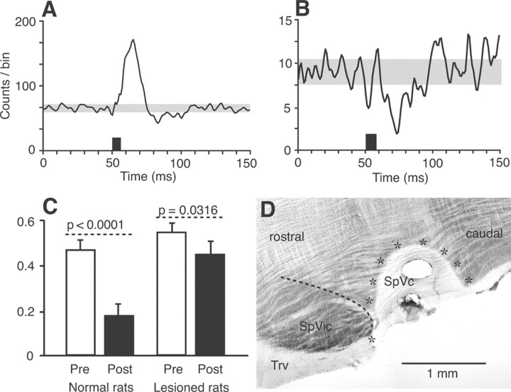
Figure 4.
Lesion of the SpVc significantly depresses the inhibitory response induced by motor cortex stimulation in the SpVi. Motor cortex-induced excitation persists after the lesion, but inhibition is markedly reduced (population PSTH of 35 multiwhisker units in A; compare with PSTH in Fig. 1B). The amount of inhibition is also reduced in monowhisker cells (population PSTH of 7 units in B). Histograms in C show the decrease in firing probability within a poststimulus time window of 20 ms (Post; 25–45 ms after the onset of cortical stimulation; black bars) with respect to firing probability in a prestimulus time window of 50 ms (Pre; white bars). Although the amount of inhibition was statistically significant in both normal and lesioned rats (59.44 and 21.47%, respectively), it was markedly decreased after lesion of the SpVc. Error bars indicate ± 1 SD. The horizontal section of the brainstem in D shows the extent of an SpVc lesion (area surrounded by stars). The dashed line outlines the border between the SpVi and SpVc. Trv, Trigeminal tract.

