
Figure 3.
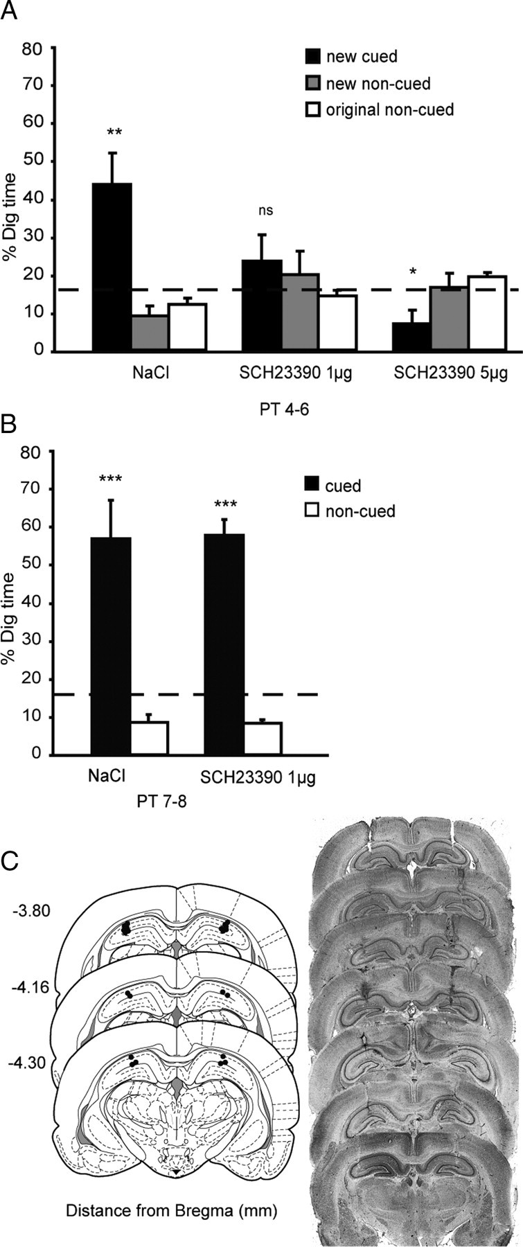
Experiment 1: A, Dopamine dependency of encoding of new paired associates. Percentage dig time in all three conditions (NaCl, SCH23390 1 μg, and SCH23390 5 μg) across the three counterbalanced probe tests (PT4–PT6). B, SCH23390 and previously trained memories. Percentage dig time for the original paired associates in two conditions (NaCl and SCH23390 1 μg). C, The left shows plots of the locations of cannulae tips (n = 11 per HPC; experiment 1). Infusion sites are marked on the appropriate section of a stereotaxic brain atlas (Paxinos and Watson, 1998). The right shows Nissl-stained sections showing examples of representative cannulae tracks in the dorsal hippocampus in each hemisphere of the brain. ns, Nonsignificant. *p < 0.05; **p < 0.01; ***p < 0.001.