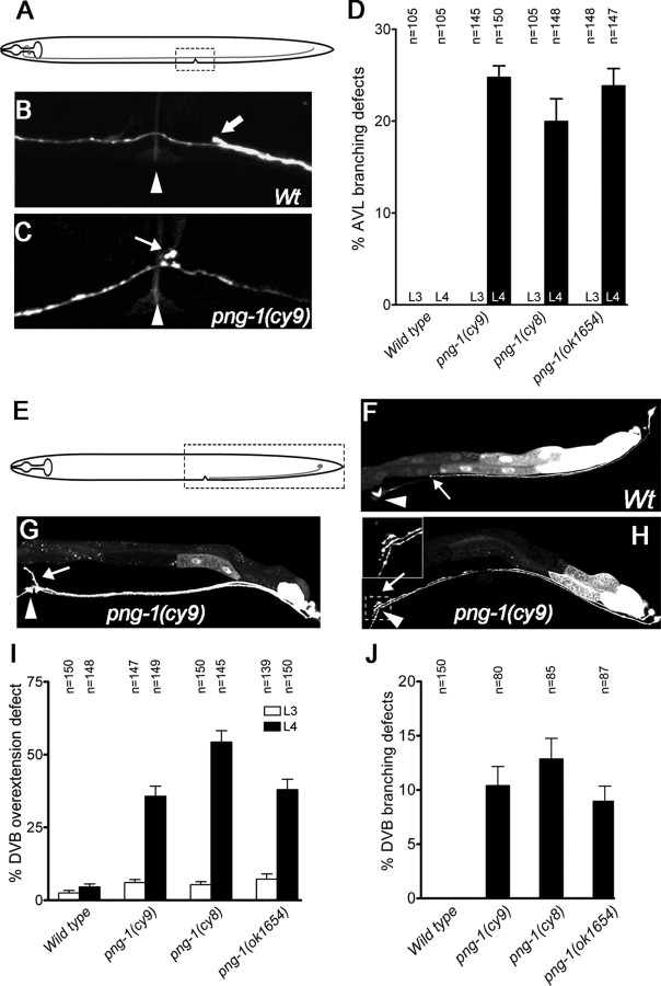
Figure 4.
Several neurons display excessive or inappropriate axonal branching at the vulva in png-1 mutants. A, E, Schematics of AVL and DVB neurons with imaged areas boxed. B, C, An AVL axon, visualized in a Punc-25::GFP; unc-30(e191) background, running adjacent to the vulva (arrowhead) in a wild type (B; Wt) and displaying an inappropriate branch (arrow) in a png-1(cy9) adult (C). Thick arrow (B) points to a DVB axon. D, Inappropriate AVL branches are observed at mid-to-late L4 stage at the vulva but not before vulval organogenesis at the L3 stage in png-1 mutants. F–H, A DVB axon visualized using an Pflp-10::GFP reporter terminating (arrow) posterior to the vulva (arrowhead) in a wild type (F), overextending anteriorly and dorsally along the vulval epithelium (arrow) (G) and displaying a vulval branch (arrow) in png-1(cy9) adults (H). Pflp-10::GFP is also variably expressed in the VulD vulval cells (arrowhead in H). I, J, DVB axon overextension defects are more severe at the mid-to-late L4 stage compared with the L3 stage in png-1 mutants (I). A subset of DVB axons that display overextension defects also branch at the vulva (J). Error bars denote SEM. Scale bars: B, C, 10 μm; F–H, 20 μm.

