
Figure 1.
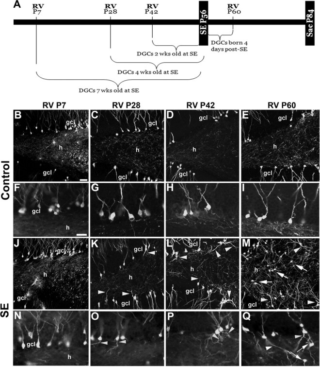
RV-GFP reporter labeling of DGCs generated at specific time points before or after SE. A, Timeline for RV labeling of newborn cells in the dentate gyrus born before or after SE. B–Q, Images of GFP-immunoreactive cells after injection of RV at the designated postnatal ages (P7, P28, P42, or P60) in rats receiving saline (B–I) or pilocarpine (J–Q) at P56. Higher magnification views of RV-GFP-labeled DGCs show granule cells with hilar basal dendrites (arrowheads) or hilar ectopic DGCs (arrows) only at specific RV labeling time points (P28, P42, or P60) with respect to SE at P56. ml, Molecular layer; gcl, granule cell layer; h, hilus. Scale bars: (in B) B–E, J–M, 50 μm; (in F) F–I, N–Q, 25 μm.