Figure 7.
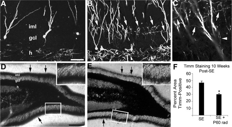
Cells born after SE contribute to MFS 10 weeks after pilocarpine treatment. A, GFP immunolabeling of a control injected with RV-GFP 4 d after saline treatment and killed 10 weeks later shows axonal labeling in the hilus (h) but only GFP+ dendrites in the inner molecular layer (iml). B, C, At 10 weeks after SE, animals that received pilocarpine and then RV-GFP 4 d later display many GFP+ axonal processes in the iml (arrows) that appear identical with those seen in the granule cell layer (gcl) and hilus (arrowheads). D, E, Denser iml Timm staining is seen at 10 weeks after SE in a sham-irradiated control (D) than in a rat irradiated beginning 4 d after pilocarpine treatment (E). F, Densitometric analysis of the percentage area of inferior blade gcl and molecular layer that is Timm stain-positive. A dose of 6 Gy fractionated irradiation administered 4 and 6 d after SE significantly decreased the amount of Timm staining (*p < 0.005). Scale bars: (in A) A–C, 50 μm; (in D) D, E, 200 μm; insets, 50 μm. Error bars indicate SEM.

