
Figure 1.
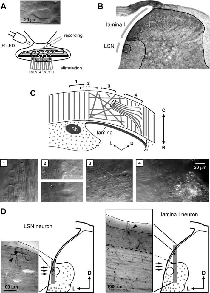
Identification of lamina I and LSN neurons in the isolated spinal cord. A, Preparation of the lumbar spinal cord with unilateral six dorsal roots, L1–L6. The roots were stimulated through suction electrodes. Inset, Lamina I neuron viewed using oblique LED illumination. B, A cross section of the fixed spinal cord (L4, 27-d-old rat) with indications of the LSN and the lamina I region accessible for the recording. Continuous lines show the border of the gray matter and the LSN. C, Schematic drawing of the fiber orientation in the dorsal and dorsolateral white matter used for the identification of the LSN and lamina I. The photographs shown below were taken from the regions (1–4) indicated on the schematic drawing. The focal plane was chosen to show the fibers. Region 1, The LSN: large neuronal cell bodies are surrounded by the parallel rostrocaudal myelinated fibers of the dorsolateral funiculus. Region 2, Transitional zone between the dorsolateral funiculus (left) and lamina I (right). Region 3, Network of the randomly oriented fibers and the scattered cell bodies in lamina I. Region 4, The medial border of accessible part of lamina I (left) and myelinated fibers in the dorsal root entry zone (right). D, Parasagittal sections (100 μm thick) of the spinal cord with a biocytin-labeled LSN neuron (left) and lamina I neuron (right). Locations of the sections are shown in the schematic drawings where asterisks indicate the cell body positions. Left, LSN neuron is found outside the spinal gray matter within the dorsolateral funiculus. Continuous lines show the dorsal borders of the white matter in the top and bottom of the section. Right, Lamina I neuron is seen within the dorsolateral gray matter. Dashed line shows the border between the gray and white matter. Continuous lines indicate the dorsal borders of the white matter in the top and bottom of the section.