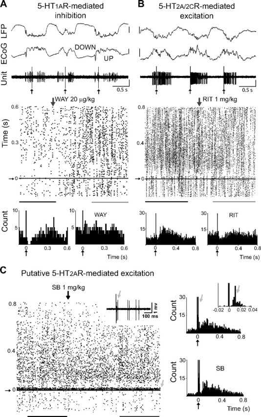
Figure 5.

Opposite modulation of FSi activity in vivo through 5-HT1ARs and 5-HT2ARs. A, B, Top, DRN stimulation (1 Hz) delivered during UP states induced transient inactivations in many FSi recorded (left), while a subset of FSi were activated (right). Vertical bar is 0.5 mV. Bottom, Raster plots and corresponding peristimulus histograms of the units shown above. Inhibitions were mediated by 5-HT1ARs and activations by 5-HT2A/2CRs. Zero is time of DRN stimulation. Peristimulus histograms were built during baseline (black lines under raster plots) and after the administration of WAY or RIT (gray lines). Lines are 5 min epochs. Vertical arrows point to the start of the injections. Small vertical arrows point to stimulus artifacts. Both units were confirmed to be PV positive. C, Raster plot and peristimulus histogram of a FSi activated by DRN stimulation during baseline (black line), and after the administration of SB (gray line). SB failed to reverse the activation (up to 2 mg/kg), suggesting that it was 5-HT2AR-mediated. Lines are 3 min epochs. Inset, Note the presence of a short-delay short-duration excitation (light gray arrows) with variable latencies. This cell was one of the few FSi recorded in the medial PFC (see Materials and Methods). Because the medial PFC projects densely to the DRN, this response may be due to feedforward inputs from cortico-raphe pyramidal neurons activated antidromically from the DRN (Puig et al., 2008). Alternatively, a glutamatergic response induced by 5-HT/glutamate corelease from serotonergic terminals should also be considered (Varga et al., 2009). This unit was confirmed to be PV positive.
