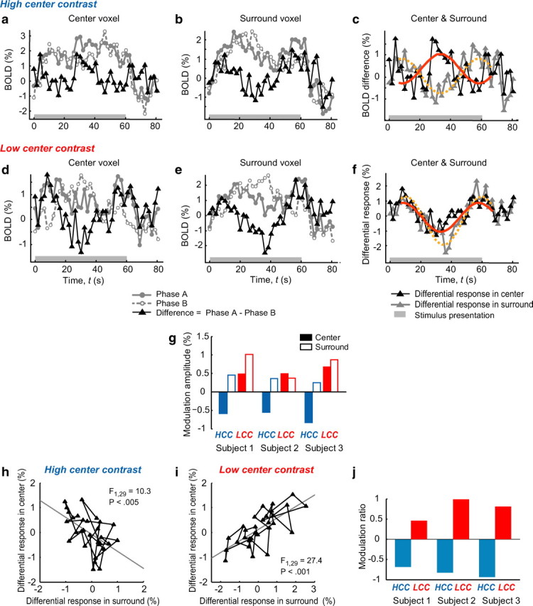
Figure 2.

The effects of contextual surround on cortical responses. a–c, Voxel- and block-averaged BOLD responses in the HCC condition from a representative subject (subject 1). a, The average differential response of voxels in the center region (black curve), which was the difference between the two original responses obtained in phase A (gray solid curve) and phase B (gray dashed curve). b, The responses of voxels in the surround. The two black curves in a and b are overlaid in c. The red solid and orange dotted curves are the sine wave fits to the differential responses in the center and surround, respectively. d–f, Similar results as shown in a–c, but for the LCC condition. g, The modulation amplitudes obtained from all three subjects. The blue and red bars are respectively for HCC and LCC conditions. The filled bars indicate the responses in the center and the open bars show the responses in the surround (excluding the center region). The bar length reflects the average of corrected modulation amplitudes from the sine wave fits and the polarity of a bar indicates whether the modulation was positively or negatively correlated with the contrast modulation in the surround (see Materials and Methods). h, i, Correlations between the responses in the center and surround in HCC (h) and LCC (i) conditions from subject 1. The time points were sampled from the duration of the test blocks, taking the hemodynamic response delay (6.06 s or 3 TRs) into consideration. The gray line in each plot is the result of the linear regression. j, The modulation ratios, defined by the slope of the linear regression shown in h or i, for the three subjects. The blue and red bars represent the modulation ratios in HCC and LCC conditions, respectively. A negative modulation ratio indicates that the contrast modulation in the surround had a suppressive effect on responses of voxels in the center region.
